Disulfide bond reduction corresponds to dimerization and hydrophobi-city changes of Clostridium botulinum type A neurotoxin
Introduction
Botulinum neurotoxins (BoNT) are potent toxins and are also therapeutic agents[1]. These botulism-causing protein toxins are produced by the anaerobic eubacterium, Clostri-dium botulinum. There are seven serologically and genetically different BoNT, named BoNT/A-G, produced by serotypes A, B, C, D, E, F, and G Clostridium botulinum strains[2]. BoNT/A is synthesized by serotype A Clostridium botulinum strain as a single-chain protein with a molecular mass of approximately 150 kDa[3,4]. This protein is post-translationally proteolyzed to form a di-chain molecule in which the two chains, a ~50 kDa light chain and a ~100 kDa heavy chain, remain linked by a disulfide bond and non-covalent bonds[5,6]. The crystal structure of BoNT/A shows a linear arrangement of three functional domains named the receptor binding domain, translocation domain, and catalytic domain[7]. These structural characters correspond to the intoxication process of BoNT/A and the mechanism of BoNT/A intoxication consisting of 4-steps: receptor binding, internalization, translocation, and cleavage of the synaptosomal associated protein of 25 kDa (SNAP-25). The receptor binding domain specifically binds to its neuronal receptor, ganglioside GT1b or GD1a, and unidentified protein receptors[8]. After receptor binding and receptor-mediated endocytosis of the neuro-toxins, they enter acidic neuronal organelles, synaptic vesicles, or endosomes[8,9]. It is believed that the translocation domain undergoes a conformational change and forms a protein-conducting channel on the membrane of the endosome to translocate the catalytic domain into the cytoplasm of neuronal cells[10]. The catalytic domain of BoNT/A is a zinc protease[11,12] and is highly specific for the C-terminus of SNAP-25, a soluble NSF accessory protein receptor (SNARE) protein complex component[13]. Cleavage of the C-terminus of SNAP-25 by the catalytic domain of BoNT/A inhibits SNARE complex formation as well as neurotransmitter release[13,14]. Inhibition of the neurotransmitter, eg, acetylcholine, released in the neuromuscular junction ultimately leads to paralysis and causes botulism[1]. This specific action at the neuromuscular junction of BoNT is increasingly being used to treat various neuromuscular disorders such as strabismus, torticollis, and blepharospasm[15,16].
Among the intoxication processes of BoNT/A and other types of BoNT, the translocation process is the least understood[1,7]. Previous results show that BoNT can form ion channels in artificial planar lipid bilayers under acidic conditions[10,17,18] and PC12 cell membrane[19]. But, how can such a big soluble protein dramatically change to a hydrophobic membrane protein? And how BoNT responds to acidic conditions to form an ion channel and how the light chain translocates across the membrane barrier is not clear. Furthermore, the quaternary structure of BoNT/A in aqueous solution is not fully resolved. Native gel electrophoresis had showed that the possible quaternary structure of BoNT/A is trimer or tetramer[20,21]. However, the crystal structure of BoNT/A meant that it was initially recognized as a dimeric species[22], but it is now recognized as a monomeric species[23]. It is believed that the quaternary structure derived from X-ray crystallography is not reliable, because under crystallization conditions the quaternary structure may change[24,25]. Thus, a clear solution to the structure of BoNT/A in solution is critical, not only because of these conflicting reports but also because it can clarify the translocation process of BoNT/A. In this study, sucrose density gradient centrifugation and chemical crosslinking experiments illustrated that only after disulfide bond reduction did BoNT/A undergo dimerization in acidic conditions. And Triton-X 114 phase-separation experiments showed that acidic conditions and disulfide reduction are factors mediating the change in hydrophobicity of BoNT/A. These results thus imply that disulfide reduction is the structural factor that corresponds to BoNT/A translocation in synaptic vesicles or endosomes.
Materials and methods
Preparation of BoNT/A neurotoxin and toxoid We purified BoNT/A neurotoxin from cultures of C botulinum type A (ATCC 7948) according to the method developed by DasGupta and Sathyamoorthy[3]. The purity was examined by 8% SDS-PAGE and Coomassie blue staining (Figure 1A). The different batch preparations contained the most nicked BoNT/A and traceable un-nicked BoNT/A. To generate antibodies against the BoNT/A toxin, we prepared the BoNT/A toxoid following the method developed by Kozaki and Sakaguchi[26]. BoNT/A (0.2 mg/mL) was toxoided by dialysis against 0.4% formalin in 0.1 mol/L phosphate buffer (pH 7.0) for 6 d at room temperature.
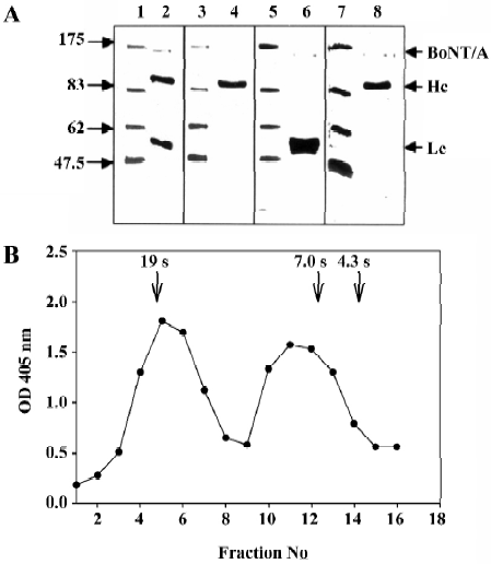
Production of antibodies The toxoid was mixed with an equal volume of Freund’s complete adjuvant. Three 0.5-mL doses of the toxoid emulsion were injected into two rabbits (2.7 and 3.2 kg) at 7-day intervals. Six weeks after the third injection, two booster injections were given. Two weeks after the booster, the rabbits were exsanguinated from the cervical artery. Polyclonal antibodies were purified from the rabbit’s serum by protein A Sepharose 4 fast-flow affinity chromatography for IgG antibodies[27]. To establish hybridoma cell lines that produce BoNT/A monoclonal antibodies, BALB/c mice were immunized intraperitoneally with 250 µg toxoid in Freund’s complete adjuvant and were boosted twice with 100 µg toxoid in Freund’s incomplete adjuvant at intervals of 2 weeks. Mice were boosted intraperitoneally with 50 µg toxoid in PBS 1 month later, and spleen cells were fused with murine plasmacytoma FO cells for 4 d by the method of Galfre and Milstein[28]. An enzyme-linked immunosorbent assay (ELISA) was used to screen culture supernatants showing positive binding to BoNT/A proteins. Hybridomas from positive-binding wells were cloned by the limiting dilution method. To obtain monoclonal antibodies, approximately 5×10⁶ hybridoma cells were inoculated into the peritoneal cavity of Pristance primed BALB/c mice, and ascitic fluid was collected on d 7–14. Monoclonal antibodies were then purified from the ascitic fluid by 40% saturated ammonium sulfate precipitation and protein A Sepharose 4 fast-flow affinity chromatography for IgG antibodies.
ELISA assay of BoNT/A The modified ELISA method developed by Engvall and Perlman[29] was used to analyze the antibodies against BoNT/A. Microtiter plates (PolySorp surface,Nalge Nunc International, Naperville, IL, USA) were coated with 100 µL per well of BoNT/A protein (0.1 mg/mL) in 0.01 mol/L PBS, pH 7.0, overnight. After blocking with 1% BSA (bovine serum albumin, Sigma Chemical Co, St Louis, MO. USA) in phosphate-buffered saline (PBS) for 1 h, plates were washed, and diluted polyclonal antibodies or hybridoma supernatant (100 µL) were added to the wells and then incubated for 1 h. Plates were next washed and incubated with alkaline phosphate (AP)-conjugated secondary antibodies for 1 h. Finally, the alkaline phosphate enzyme activity was developed with the addition of substrate, and readings were taken at an optical density (OD) of 405 nm with a Dynatech MR700 microplate reader. For the BoNT/A immunoassay, a sandwich ELISA was developed. The ELISA plate wells were coated with 0.1 µg (1 µg/mL) monoclonal antibodies overnight and then incubated with 100 µL of the BoNT/A protein or BoNT/E (0.007 to 1 µg/mL) or samples from sucrose density gradient centrifugation fractions for 1 h. The bound BoNT/A proteins were then labeled with 100 µL polyclonal antibodies (1 µg/mL), probed with AP-conjugated goat anti-rabbit IgG, and quantified using the colorimetric assay. Absorbance at 405 nm was measured with an ELISA plate reader.
SDS-PAGE and immunoblotting Proteins were separated by SDS-PAGE according to the procedure of Laemmli[30] or the method of Weber and Osborn[31] on a mini ProteinII system (Bio-Rad, Hercules, CA, USA). After SDS-PAGE fractionation, the proteins were electrotransferred to a polyvinyli-dene difluoride (PVDF) membrane (Millipore, Bedford, MA, USA). The resulting membrane was blocked with PBS containing 0.05% Tween-20 and 5% BSA (Sigma) at room temperature. Subsequently, the membrane was incubated with primary antibody in PBS at 4 °C overnight and then with AP- or horseradish peroxidase (HRP)-conjugated secondary antibodies (Jackson ImmuneResearch, West Grove, PA, USA). The AP on the membrane was detected using a Western-blue kit (Promega, Madison, WI, USA), and an enhanced chemiluminescence kit (Pierce, Rockford, USA) was used to detect the HRP on the membrane.
Sucrose density gradient centrifugation An SW-41 rotor (Beckman, Palo Alto. CA, USA) was used to perform the sucrose density gradient centrifugation experiments. Sucrose density gradients in 0.1 mol/L HEPES buffer (Sigma) at different pH values (pH 7.5 and 4.5) were prepared with a sucrose gradient maker from two sucrose stock solutions containing 5% and 20% (w/v) sucrose (Merck-Shuchardt Chemical Co, Germany). Gradients were maintained at 4 °C cold room for 4–6 h before use. Samples were then applied on top of the gradients, and were centrifuged at 5 °C for 19 h. Each sucrose density gradient centrifugation experiment consisted of a calibration control containing the marker proteins (Sigma), thyroglobulin (19 S, 669 kDa), catalase (11.3 S, 232 kDa, purchased from Pharmacia Upjohn, Peapack, NJ, USA), alcohol dehydrogenase (7.0 S, 150 kDa), and BSA (4.3 S, 66 kDa). Following centrifugation, sample fractions (0.65 mL) were removed from the bottom of the centrifuge tube for the ELISA of BoNT/A.
Chemical crosslinking A homobifunctional crosslinking reagent, Bis (sulfosuccinimidyl) suberate (BS3, Pierce), was used in this study. The functional group of BS3 is a sulfo-NHS ester, which forms covalent linkages with the neighboring primary amines. An 11.4-Å-long linker separates the functional groups of BS3.
Prior to performing the crosslinking reactions, purified BoNT/A proteins (200 µg/mL) were dialyzed against the buffer solution containing 50 mmol/L HEPES and 100 mmol/L NaCl at pH 7.5, 6.1, and 4.5, respectively. For the reduced BoNT/A, the toxins were treated with 20 mmol/L DTT at 37 °C for 30 min before the crosslinking reactions. For crosslinking BoNT/A with BS3, 4 µL of freshly prepared 7.5 mmol/L B3 was added to 20 µL of purified reduced or non-reduced toxin (2 µg) and incubated at room temperature. Aliquots of freshly prepared 7.5 mmol/L BS3 (3 µL) were added to the reaction mixtures 20 and 60 min later. Reactions were stopped 1 h after the final addition of BS3 by adding 50 mmol/L glycine. After the crosslinking reactions, these samples were subjected to SDS-PAGE fractionation and assayed by Western blotting with BoNT/A polyclonal antibodies.
Triton-X 114 phase partition Purified BoNT/A proteins (200 µg/mL) were dialyzed against the buffer solution containing 50 mmol/L HEPES and 100 mmol/L NaCl at pH 7.5, 6.1, and 4.5, respectively. For the reduced BoNT/A, the toxins were treated with 20 mmol/L DTT at 37 °C for 30 min before the phase-separation experiment.
The Triton X-114 phase-separation experiment was carried out according to the method of Bordier (1981)[32]. A Triton X-114 (10%, 20 µL) solution was added to the BoNT/A protein toxins (180 µL) and incubated on ice for 5 min and then incubated at 30 °C for 5 min. After centrifuging for 5 min in an Eppendorf centrifuge, 10% Triton X-114 (10 µL) was added to the water phase, and 0.1 mol/L HEPES buffer was added to 200 µL, producing a final Triton X-114 concentration of 0.5%. Triton X-114 phase separation was repeated again, and the detergent phase was added together. Triton X-114 was again added to the water phase to obtain a final concentration of 2%. After phase separation, the water phase was removed to a new Eppendorf tube, and the detergent phase was discarded. Then 0.1 mol/L HEPES buffer was added to the detergent phase and water phase solution to 500 µL, respectively. Aliquots of the detergent phase and water phase samples (40 µL) were added to 15 µL of the SDS-PAGE sample solution. These samples were analyzed by Western blotting.
1-Anilino-8-naphthalene sulfonate (ANS) binding ANS fluorescence measurements were made on a Perkin-Elmer spectrofluorometer model LS-50B. ANS (Molecular Probes, Eugene, OR, USA) was dissolved in absolute ethanol, and its concentration was determined by spectrophotometer [ε372(ANS) = 8000 mol-1cm-1]. Purified BoNT/A proteins (20 µg/mL) were dialyzed against the buffer solution containing 50 mmol/L HEPES and 100 mmol/L NaCl at pH 4.5. For the reduced BoNT/A, the toxins were treated with 20 mmol/L DTT at 37 °C for 30 min before the ANS binding experiment. One microliter aliquots of 5 mmol/L ANS were added into a 1-cm path length cuvette containing 1 mL of 0.125 µmol/L proteins. BoNT/A or reduced BoNT/A with ANS was then excited at 360 nm, and the emission was recorded between 400 and 580 nm. The excitation and emission slit widths were both set to 15 nm. All measurements were made at 25 °C.
Results
Development of the immunoassay of BoNT/A Because BoNT/A is a zinc protease[11,12], its biochemical characterization is not convenient. To facilitate the biochemical analysis of BoNT/A, we tried to establish an immunoassay for BoNT/A. We identified two monoclonal antibodies, named H-4F and H-3, that specifically recognize the heavy chain of BoNT/A, and one monoclonal antibody, L-1H, that specifically recognizes the light chain of BoNT/A (Figure 1A). These three monoclonal antibodies then served as capture antibodies for the ELISA of BoNT/A. We found that L-1H failed to act as a capture antibody for the ELISA of BoNT/A when combined with the polyclonal antibodies generated from the rabbit. However, the H-4F and H-3 monoclonal antibodies could serve as capture antibodies and detected BoNT/A at as low as 10 ng/mL BoNT/A by sandwich ELISA. Further-more, this assay can distinguish BoNT/E and BoNT/A, even though the presence of BoNT/E was as high as 100 ng/mL (data not shown). Thus, we performed the ELISA to characterize the crude extract from cultures of C botulinum type A after sucrose density gradient centrifugation. It is well established that in addition to a 150-kDa toxic protein com-ponent, a progenitor toxin with a molecular mass of 900 kDa, composed of hemagglutinin proteins and a nontoxic non-hemagglutinin protein, also exists in the crude extract of cultures of C botulinum type A[8]. Figure 1B shows that the crude extract existed as two forms of BoNT/A, 7S and 19S, which correspond to the pure BoNT/A toxin protein and the 900-kDa BoNT/A complex protein. These results indicated that ELISA can serve as a convenient tool to assay the biochemical properties of BoNT/A.
Sucrose density gradient centrifugation demonstrated that disulfide reduction is necessary for the dimer formation of BoNT/A under acidic conditions Clostridium botulinum neurotoxin, like tetanus toxin and diphtheria toxin, forms ion channels in planar lipid bilayers under acidic conditions[10,16,17]. The ion channel activity had been proposed as the translocation mechanism of BoNT/A in neuronal cells[8]. Thus, we hypothesized that type A C botulinum neurotoxin proteins can form oligomeric proteins in acidic conditions, like the anthrax protective antigen[33]. Sucrose density gradient centrifugation combined with the ELISA was used to analyze the oligomer states of BoNT/A. Figure 2A shows that the purified BoNT/A contained the toxin component only, without nontoxic protein contamination. These nontoxic protein contaminations may be associated with BoNT/A and may interfere with judgment concerning the oligomeric state of BoNT/A. After dialysis of the purified BoNT/A in pH 4.5 and 7.5 HEPES buffer, 0.2 mg/mL BoNT/A was loaded on a 5%–20% sucrose density gradient and centrifuged at 151 200×g for 19 h at 5 °C. We found that the BoNT/A protein migrated as a 7S, monomeric toxin with a molecular mass of about 150 kDa, in both neutral and acidic conditions (Figure 2B). These results suggest that factors other than an acidic condition are necessary for the oligomerization of BoNT/A.
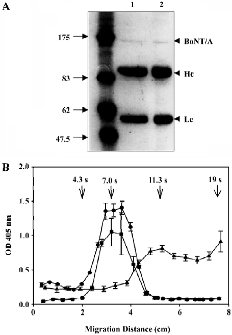
Previous studies showed that the heavy chain of BoNT/B, but not the entire toxin, can form ion channels under acidic conditions[10]. Recent results also indicated, after DTT reduction, that the BoNT/A structure is more flexible than the non-reduced form[34]. Thus, we assumed that reduction of the disulfide bond between the heavy chain and the light chain may facilitate oligomeric channel formation of BoNT/A. To test this assumption, BoNT/A proteins were treated with DTT before being subjected to sucrose density gradient centrifugation at pH 4.5. We found that treatment of BoNT/A with 20 mmol/L DTT at 37 °C for 30 min efficiently reduced the disulfide bond between the heavy and light chains (compare lanes 7 of Figure 4A and 4B). Figure 2B shows that the DTT-reduced BoNT/A proteins migrated as a major peak at the 12S position with an approximate molecular weight of 300 kDa. This result suggests that BoNT/A proteins can form dimers under acidic conditions only after disulfide bond reduction.
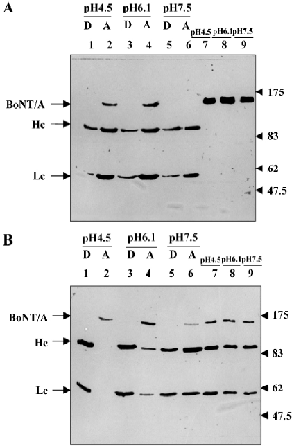
Chemical crosslinking studies of type A Clostridium botulinum neurotoxin The channel stoichiometry formed by the BoNT has not been fully understood, but a dimer and tetramer structure has been proposed[10,17,18,21,35]. To further clarify the oligomeric state of BoNT/A, we used BS3 (0.5 mmol/L) to perform chemical crosslinking on BoNT/A after extensive dialysis in pH 4.5 and 7.5 HEPES buffer, respec-tively. Although the sucrose density gradient centrifugation experiment indicated that BoNT/A proteins exist as monomers under these conditions, the crosslinking pattern of BoNT/A showed a ladder form, from monomer to tetramer (Figure 3, lanes 2 and 3). In contrast, the crosslinking pattern of 20 mmol/L DTT-reduced BoNT/A showed that, under neutral conditions, the final product was a monomer (Figure 3, lane 4) and that in an acidic conditions the final products were dimers of BoNT/A (Figure 3, lane 5). From this result, combined with the sucrose density gradient centrifugation, we suspected that BoNT/A can form dimers after reduction, and the tetramer observed in the non-reduced BoNT/A crosslinking study may have arisen from intermolecular crosslinking artifacts.
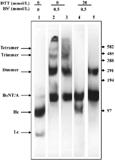
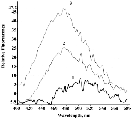
Disulfide bond reduction and acidic pH are responsible for the hydrophobic change of type A C botulinum neurotoxin Previous studies have shown that botulinum neurotoxins can form ion channels on planar lipid bilayers[10,17,18]. This suggests that aqueous BoNT/A possibly becomes a hydrophobic-like protein in order to cross the hydrophobic barrier. Interestingly, Figure 2B shows that, in addition to the dimeric BoNT/A protein, there are large BoNT/A aggregations after disulfide bond reduction in an acidic condition. This result may imply that the hydrophobicity of BoNT/A undergoes a dramatic change in acidic conditions after disulfide bond reduction. It has been shown that the Triton X-114 phase separation experiment can distinguish membrane proteins from water-soluble proteins[32]. So we first prepared the BoNT/A proteins under different pH conditions, either reduced with 20 mmol/L DTT or not, and then performed the Triton X-114 phase separation experiment. As shown in Figure 4A, most non-reduced BoNT/A proteins were still hydrophilic-like proteins. Densitometric scans indicated that less than 20% of the BoNT/A proteins were partitioned into the detergent phase (Figure 4A, lanes 1, 3, and 5). These results indicate that factors other than the acidic condition are necessary for the hydrophobicity change of BoNT/A. Indeed, under an acidic condition, reduced BoNT/A dramatically changed to hydrophobic proteins (Figure 4B, compare lanes 1 and 2 and lanes 3 and 4). A densitometric scan indicated that more than 90% of the heavy chains and light chains of BoNT/A were partitioned into the Triton-X114 phase. However, the disulfide bond-reduced BoNT/A was still a hydrophilic-like protein under a neutral condition, and less than 40% of the heavy chains and light chains of BoNT/A were partitioned into the Triton-X114 phase (Figure 4B, compare lanes 5 and 6). To further confirm the results of Triton X-114 phase separation, we probed the hydrophobic change in BoNT/A with a hydrophobic-sensitive dye, ANS. The ANS anion is an often-utilized hydrophobic probe for proteins[36]. When excited at 360 nm, the binding of nonpolar ANS to the hydrophobic region of a protein is associated with enhanced ANS fluorescence emission spectra at 480 nm. We found that no obvious ANS fluorescence was observed when non-reduced BoNT/A at pH 7.5 (Figure 5, line 1). However, there was nearly a two-fold increase in the fluorescence intensity when ANS was bound to the reduced BoNT/A than to the non-reduced BoNT/A at pH 4.5 (Figure 5, compare lines 2 and 3). Combining the Triton X-114 phase separation and ANS binding experiments, our observations imply that an acidic condition and disulfide reduction are both necessary for the change in hydrophobicity of BoNT/A.
Discussion
As to their intracellular actions, many plant and bacterial toxins do not directly penetrate the plasma membrane but go through a low-pH compartment (ie, synaptic vesicle or endosome)[37]. These toxins have evolved sophisticated mechanisms to translocate their active portion through the hydrophobic barrier of these acidic organelles. For example, some studies have shown that the diphtheria toxin undergoes a conformational change and exposes hydrophobic sites within the acidic endosomal lumen[37]. The exposure of hydrophobic sites then induces membrane insertion and channel formation, and results in translocation of the catalytic domain. These results indicate that an acidic pH is the main factor triggering membrane insertion of diphtheria toxin, but disulfide bond reduction and nicking of the diphtheria toxin are not necessary for membrane insertion[38]. Similarly, there are experiments which show that an acidic pH is necessary for membrane insertion and ion channel formation of BoNT[8,10]. However, circular dichroism and fluorescence spectroscopic studies revealed that the conformation of BoNT/A does not undergo a drastic change over a range of pH 6–9[39], and near-UV circular dichroism spectroscopy indicated that the structural features of BoNT/A change considerably upon disulfide reduction[34]. So, disulfide reduction might influence the membrane insertion of BoNT. Consistent with these spectroscopic studies, Triton X-114 phase separation showed that disulfide bond reduction and an acidic condition (pH 4.5 and 6) were both required for the increase in hydrophobicity of BoNT/A protein molecules (Figure 4). In addition, the large aggregate found in sucrose density gradient centrifugation may reflect the hydrophobic change of the disulfide bond-reduced BoNT/A protein in an acidic condition (Figure 2B), like the diphtheria toxin and protective antigen of anthrax toxin found in acidic conditions[38,40]. Interestingly, the traceable un-nicked BoNT/A did not change its hydrophobicity even after disulfide reduction (Figure 4B, lanes 2, 4, and 6). This is similar to the anthrax toxin protective antigen in which low pH-induced proteolytically nicked PA63 hydrophobicity increases but does not induce a change in hydrophobicity in the intact protective antigen[40]. Consistent with these observations, we observed that the enhancement of fluorescence of ANS by the disulfide-reduced BoNT/A was nearly two-fold stronger than that of the non-reduced form in an acidic condition (Figure 5) and Kamata et al also recently showed that botulinum type B-nicked neurotoxin becomes hydrophobic more quickly and extensively than does the un-nicked toxin under acidic pH conditions[41]. These results suggest that the nick between the heavy chain and the light chain may also involve membrane insertion of botulinum neurotoxins in acidic pH conditions.
The channel stoichiometry of BoNT/A was studied by sucrose density gradient centrifugation and chemical crosslinking experiments. Sucrose density gradient centrifugation showed that the non-reduced BoNT/A did not form oligomers even in an acidic pH (Figure 2B). This result is consistent with the light-scattering experiment, without the hydraulic pressure in the sucrose density gradient centrifugation, which revealed that only the monomer of BoNT/A exists in solution over a broad pH range[42]. But, in the chemical crosslinking study, tetramers emerged as the final product of BS3 crosslinked, non-reduced BoNT/A (Figure 3, lanes 2 and 3). There are two possible reasons to explain these contrasting results. First, the tetramer observed in BS3 crosslinked BoNT/A is intermolecular crosslinking artifacts and not intramolecular crosslinking under the crosslinking condition. Second, the interaction between the tetramer of BoNT/A is weak, and the hydraulic pressure during the sucrose density gradient centrifugation is large enough to dissociate the tetramer into monomers.
Furthermore, we also found that reduced BoNT/A proteins can form dimers under acidic conditions, either by sucrose density gradient centrifugation (Figure 2B) or chemical crosslinking (Figure 3, lanes 5). Recently, Cai and Singh had reported that BoNT/A, by native gel electrophoresis, chemical crosslinking, gel filtration and fluorescence anisotropy but not by sucrose density gradient centrifugation, exists as a dimer in both reduced and non-reduced conditions[43]. Interestingly, using fluorescence anisotropy they also found that a high concentration (above 200 nmol/L) of BoNT/A existed as a dimer, while at a low concentration (20 nmol/L) it existed as a monomer. Comparing with the results of Cai and Singh, we also found that BoNT/A (approximately 440 nmol/L) can exist as a dimer, trimer or tetramer by BS3 crosslinking experiments (Figure 3, lanes 2 and 3). This result may imply that there are weak interaction between the non-reduced BoNT/A and this may change the equilibrium between oligomer states at neutral pH condition. But the hydraulic pressure during centrifugation may affect the weak interactions and only monomer was observed in the sucrose density gradient centrifugation experiment (Figure 2B). From these observations, we concluded that BoNT/A forms a dimer structure after disulfide reduction in acidic conditions, implying that the BoNT/A channel stoichiometry is a dimer. Although, molecular modeling of the pore structure of BoNT/A and electric density mapping of the BoNT/A protein both suggest a tetramer structure for BoNT/A[18,34]. This result is consistent with reports by Donovan and Middlebrook, who measured the dependence of conductance on BoNT/C concentration and also indicated that the stoichiometry of the channel is a dimer[17].
Recently, a triterpenoid derivative, toosendanin, extracted from the bark of Melia toosendan Sieb et Zucc had been demonstrated with antibotulismic effect via interference with toxin translocation[44,45]. In this study, we show that disulfide reduction is responsible for the change in hydrophobicity and dimer formation of BoNT/A in an acidic condition. This finding implies that compounds that block this disulfide bond reduction may serve as potential therapeutic agents for botulism. Furthermore, mutations on disulfide reduction sites might reduce the toxicity of BoNT by preventing the translocation process. These translocation-deficient mutated BoNT may serve as good vaccine candidates for botulism.
Acknowledgement
We greatly thank Mrs Rey-fun SHEU for technical assistance.
References
- Johnson EA. Clostridial toxins as therapeutic agents: benefits of nature’s most toxic proteins. Annu Rev Microbiol. 1999;53:551-75.
- Niemann H, Blasi J, Jahn R. Clostridial neurotoxins: new tools for dissecting exocytosis. Trends Cell Biol 1994;4:179-85.
- DasGupta BR, Sathyamoorthy VS. Purification and amino acid composition of type A botulinum neurotoxin. Toxicon 1984;22:415-24.
- Krysinski EP, Sugiyama H. Nature of intracellular type A botulinum neurotoxin. Appl Environ Microbiol 1981;41:675-8.
- DasGupta BR, Sathyamoorthy VS. Separation, purification, partial characterization and comparison of the heavy and light chains of botulinum neurotoxin types A, B, and E. J Biol Chem 1985;260:10461-6.
- Singh BR, Li B, Read D. Botulinum versus tetanus neurotoxins: Why is botulinum neurotoxin but not tetanus neurotoxin a food poison? Toxicon 1995;33:1541-7.
- Lacy DB, Tepp W, Choen AC, DasGupta BR, Stevens RC. Crystal structure of botulinum neurotoxin type A and implications for toxicity. Nat Struct Biol 1998;5:898-902.
- Montecucco C, Schiavo G. Structure and function of tetanus and botulinum neurotoxins. Q Rev Biophys 1995;28:423-72.
- Matteoli M, Verderio C, Rossetto O, Iezzi N, Coco S, Schiavo G, et al. Synaptic vesicle endocytosis mediates the entry of tetanus neurotoxin into hippocampal neurons. Proc Natl Acad Sci USA 1996;93:13310-5.
- Hoch DH, Romero-Mira M, Erlich BE, Finkelstein A, DasGupta BR, Simpson LL. Channels formed by botulinum, tetanus, and diphtheria toxins in planar lipid bilayers: relevance to translocation of proteins across membranes. Proc Natl Acad Sci USA 1985;82:1692-6.
- Oguma K, Fujinaga Y, Inoue K. Structure and function of Clostridium botulinum toxins. Microbiol Immunol 1995;39:161-8.
- Schiavo G, Rossetto O, Catsicas S, Polverino de Laureto P, DasGupta BR, Benfenati F, et al. Identification of the nerve terminal targets of botulinum neurotoxins serotypes A, D, and E. J Biol Chem 1993;268:23784-7.
- O’Sullivan GA, Mohammed N, Foran PG, Lawrence GW, Dolly JO. Rescue of exocytosis in botulinum neurotoxin A-poisoned chromaffin cells by expression of cleavage-resistant SNAP-25. J Biol Chem 1999;274:36897-904.
- Sutton RB, Fasshauer D, Jahn R, Brunger AT. Crystal structure of a SNARE complex involved in synaptic exocytosis at 2.4Å resolution. Nature 1998;395:347-53.
- Montecucco C, Schiavo G, Tugnoli V, de Grandis D. Botulinum neurotoxins: mechanism of action and therapeutic applications. Mol Med Today 1996;2:418-24.
- Scoot AB. Botulinum injection into extraocular muscle as an alternative to strabismus surgery. Ophthalmology 1980;87:1044-9.
- Donovan JJ, Middlebrook JL. Ion-conducting channels produced by botulinum toxin in planar lipid membranes. Biochemistry 1986;25:2872-6.
- Motal MS, Blewitt R, Tomich JM, Montal M. Identification of an ion channel-forming motif in the primary structure of tetanus and botulinum neurotoxins. FEBS Lett 1992;313:12-8.
- Sheridan RE. Gating and permeability of ion channels produced by botulinum toxin types A and E in PC12 cell membranes. Toxicon 1998;36:703-17.
- Shone CC, Hambleton P, Melling J. Inactivation of Clostridium botulinum type A neurotoxin by trypsin and purification of two tryptic fragments. Proteolytic action near the COOH- terminus of the heavy subunit destroys toxin-binding activity. Eur J Biochem 1985;151:75-82.
- Ledoux DN, Be XH, Singh BR. Quaternary structure of botulinum and tetanus neurotoxins as probed by chemical cross-linking and native gel electrophoresis. Toxicon 1994;32:1095-104.
- Stevens RC, Evenson ML, Tepp W, DasGupta BR. Crystallization and preliminary X-ray analysis of botulinum neurotoxin type A. J Mol Biol 1991;222:877-80.
- Lacy DB, Tepp W, Cohen AC, DasGupta BR, Stevens RC. Crystal structure of botulinum neurotoxin type A and implications for toxicity. Nat Struct Biol 1998;5:898-902.
- Lubkowski J, Bujacz G, Boque L, Domalile DJ, Handel TM, Wlodawer A. The structure of MCP-1 in two crystal forms provides a rare example of variable quaternary interactions. Nat Struct Biol 1997;4:64-9.
- Singh BR, Fu FN, Ledoux DN. Crystal and solution structures of superantigenic staphylococcal enterotoxins compared. Nat Struct Biol 1994;1:358-60.
- Kozaki S, Sakaguchi G. Antigenicities of fragments of Clostridium botulinum type B derived toxin. Infect Immun 1975;11:932-6.
- Ey PL, Prowse SJ, Jenkin CR. Isolation of pure IgG1, IgG2a, and IgG2b immunoglobulins from mouse serum using Protein A-Sepharose. Immunochemistry 1978;15:429-36.
- Galfre G, Milstein C. Preparation of monoclonal antibodies: strategies and procedures. Meth Enzymol 1981;73:3-46.
- Engvall E, Perlman P. Enzyme linked immunosorbent assay ELISA. III. Quantitation of specific antibodies by enzyme-labeled anti-immunoglobulin in antigen-coated tubes. J Immunol 1972;109:129-35.
- Laemmli UK. Cleavage of structural proteins during the assembly of the head of bacteriophage T4. Nature 1970;227:680-5.
- Weber K, Osborn M. The reliability of molecular weight determinations by dodecyl-sulfate-polyacrylamide gel electrophoresis. J Biol Chem 1969;244:4406-12.
- Bordier C. Phase separation of integral membrane proteins in Triton X-114 solution. J Biol Chem 1981;256:1604-7.
- Milne JC, Furlong D, Hanna PC, Wall JS, Collier RJ. Anthrax protective antigen forms oligomers during intoxication of mammalian cells. J Biol Chem 1994;269:20607-12.
- Cai S, Sakar HK, Singh BR. Enhancement of the endopeptidase activity of botulinum neurotoxin by its associated proteins and dithiothreitol. Biochemistry 1999;38:6903-10.
- Schmid MF, Robinson JP, DasGupta BR. Direct visualization of botulinum neurotoxin-induced channels in phospholipid vesicles. Nature 1993;364:827-30.
- Matulis D, Baumann CG, Bloomfield VA, Lovrien RE. 1-Anilino-8-naphthalene sulfate as a protein conformational tightening agent. Biopolymer 1999;49:451-8.
- Alouf J. Source book of bacterial protein toxins. In: Montecucco C, Papini E, Schiavo G, editors. Molecular models of toxin membrane translocation. New York: Academic Press; 1991. p 45–56.
- Blewitt MG, Chung LA, London E. Effect of pH on the conformation of diphtheria toxin and its implications for membrane penetration. Biochemistry 1985;24:5458-64.
- Datta A, DasGupta BR. Circular dichroic and fluorescence spectroscopic study of the conformation of botulinum neurotoxin types A and E. Mol Cell Biochem 1988;79:153-9.
- Koehler TM, Collier RJ. Anthrax toxin protective antigen: low-pH-induced hydrophobicity and channel formation in liposomes. Mol Microbiol 1991;5:1501-6.
- Kamata Y, Tahara R, Kozaki S. Difference in hydrophobicity between botulinum type B activated and non-activated neurotoxins under low pH conditions. Toxicon 2000;38:1247-51.
- Chen F, Kuziemko GM, Stevens RC. Biophysical characterization of the stability of the 150-kilodalton botulinum toxin, the nontoxic component, and the 900-kilodalton botulinum toxin complex species. Infect Immun 1998;66:2420-5.
- Cai S, Singh BR. A correlation between differential structural features and the degree of endopeptidase activity of type A botulinum neurotoxin in aqueous solution. Biochemistry 2001;40:4693-702.
- Shi YL, Wang ZF. Cure of experimental botulism and antibotulism effect of toosendanin. Acta Pharmacol Sin 2004;25:839-48.
- Li MF, Shi YL. Toosendanin interferes with pore formation of botulinum toxin type A in PC12 cell membrane. Acta Pharmacol Sin 2005;27:66-70.
